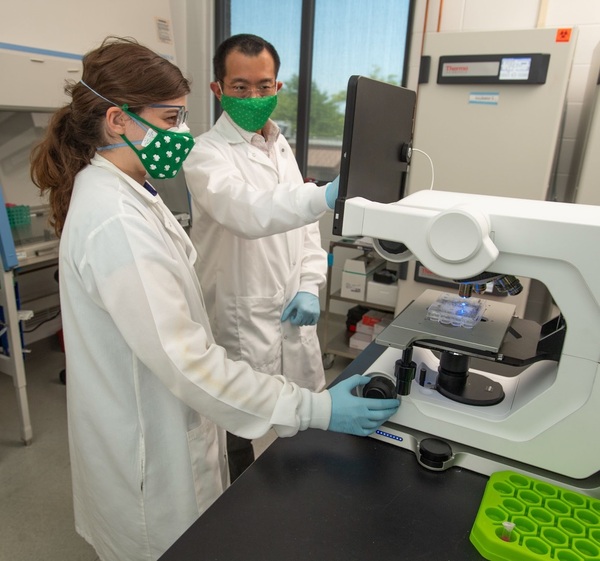
Notre Dame researchers’ “backpacks” restore damaged stem cells
Within a newborn’s umbilical cord lie potentially life-saving stem cells that can be used to fight diseases like lymphoma and leukemia. That is why many new parents elect to store (“bank”) their infant’s stem cell-rich umbilical cord blood. But in the 6-15% of pregnancies affected by gestational diabetes, parents lack this option because the condition damages the stem cells and renders them useless.
Now, in a study forthcoming in Communications Biology, bioengineers at the University of Notre Dame have shown that a new strategy can restore the damaged stem cells and enable them to grow new tissues again.
At the heart of this new approach are specially engineered nanoparticles. At just 150 nanometers in diameter—about a quarter of the size of a red blood cell—each spherical nanoparticle is able to store medicine and deliver it just to the stem cells themselves by attaching directly onto the stem cells’ surface. Due to their special formulation or “tuning,” the particles release the medicine slowly, making it highly effective even at very low doses.
Donny Hanjaya-Putra, an assistant professor of aerospace and mechanical engineering, bioengineering graduate program at Notre Dame who directs the lab where the study was conducted, describes the process using an analogy. “Each stem cell is like a soldier. It is smart and effective; it knows where to go and what to do. But the ‘soldiers’ we are working with are injured and weak. By providing them with this nanoparticle ‘backpack,’ we are giving them what they need to work effectively again.”

The main test for the new “backpack”-equipped stem cells was whether or not they could form new tissues. Hanjaya-Putra and his team tested damaged cells without “backpacks” and observed that they moved slowly and formed imperfect tissues. But when Hanjaya-Putra and his team applied “backpacks,” previously damaged stem cells began forming new blood vessels, both when inserted in synthetic polymers and when implanted under the skin of lab mice, two environments meant to simulate the conditions of the human body.
Although it may be years before this new technique reaches actual healthcare settings, Hanjaya-Putra explains that it has the clearest path of any method developed so far. “Methods that involve injecting the medicine directly into the bloodstream come with many unwanted risks and side effects,” Hanjaya-Putra explains. In addition, new methods like gene editing face a long path to Food and Drug Administration (FDA) approval. But Hanjaya-Putra’s technique used only methods and materials already approved for clinical settings by the FDA.
Hanjaya-Putra attributes the study’s success to a highly interdisciplinary group of researchers. “This was a collaboration between chemical engineering, mechanical engineering, biology, and medicine—and I always find that the best science happens at the intersection of several different fields.”
The study’s lead author was a former Notre Dame postdoctoral Loan Bui, now a faculty member at the University of Dayton in Ohio; stem cell biologist Laura S. Haneline and former postdoctoral fellow Shanique Edwards from the Indiana University School of Medicine; Notre Dame Bioengineering Ph.D. students Eva Hall and Laura Alderfer; Notre Dame undergraduates Pietro Sainaghi, Kellen Round, and 2021 valedictorian Madeline Owen; Prakash Nallathamby, research assistant professor, aerospace and mechanical engineering; and Siyuan Zhang from the UT Southwestern Medical Center.
The researchers hope their approach will be used to restore cells damaged by other types of pregnancy complications, such as preeclampsia. “Instead of discarding the stem cells,” Hanjaya-Putra says, “in the future we hope clinicians will be able to rejuvenate them and use them to regenerate the body. For example, a baby born prematurely due to preeclampsia may have to stay in the NICU with an imperfectly formed lung. We hope our technology can improve this child’s developmental outcomes.”
The study was made possible by funding from Notre Dame’s Advancing Our Vision Initiative in Stem Cell Research, Notre Dame’s Science of Wellness Initiative, the Indiana Clinical and Translational Science Institute, the American Heart Association, and the National Institutes of Health.
Find out more about how Notre Dame conducts (non-embryonic) stem cell research in accordance with Catholic ethics at https://stemcell.nd.edu/ethics/.
About Notre Dame Research:
The University of Notre Dame is a private research and teaching university inspired by its Catholic mission. Located in South Bend, Indiana, its researchers are advancing human understanding through research, scholarship, education, and creative endeavor in order to be a repository for knowledge and a powerful means for doing good in the world. For more information, please see research.nd.edu or @UNDResearch.
This article was written by Brett Beasley and originally published by Notre Dame Research on July 5, 2022. For more information, please visit their website.
July 5, 2022


